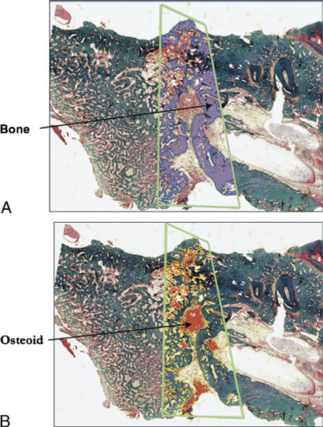
FIGURE 4.

A, Sagittal section of midportion of rat mandible stained with Gomori trichome. Green trapezoid outlines distraction regenerate. Bone is highlighted in purple by color thresholding. B, Sagittal section of midportion of rat mandible stained with Gomori trichome. Green trapezoid outlines distraction regenerate. Nonmineralized matrix is highlighted in red by color thresholding.
