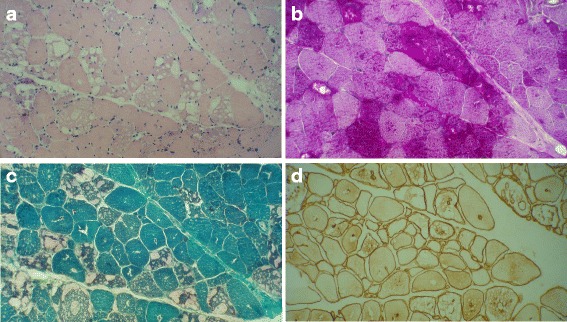
Fig. 2.

Biopsy from right vastus lateralis of a 46-year-old woman with LOPD. She had a long history of muscle weakness presenting with respiratory insufficiency. a Prominent fiber size variation and excess internalized nuclei with variable-sized subsarcolemmal and cytoplasmic vacuoles (H&E stain x200). b Pronounced vacuolation of many fibers, some vacuoles are slightly red-rimmed (modified Gomori trichrome stain x200). c Pronounced glycogen accumulation as intense staining of subsarcolemmal and cytoplasmic vacuoles (PAS stain x200). d Immunolabeling of multiple membrane-bound cytoplasmic vacuoles with dystrophin (Dystrophin 1, Biogenex Co. x200). Images provided by Dr Yalda Nilipour
