. 2015 Oct 16;4:e08961. doi: 10.7554/eLife.08961

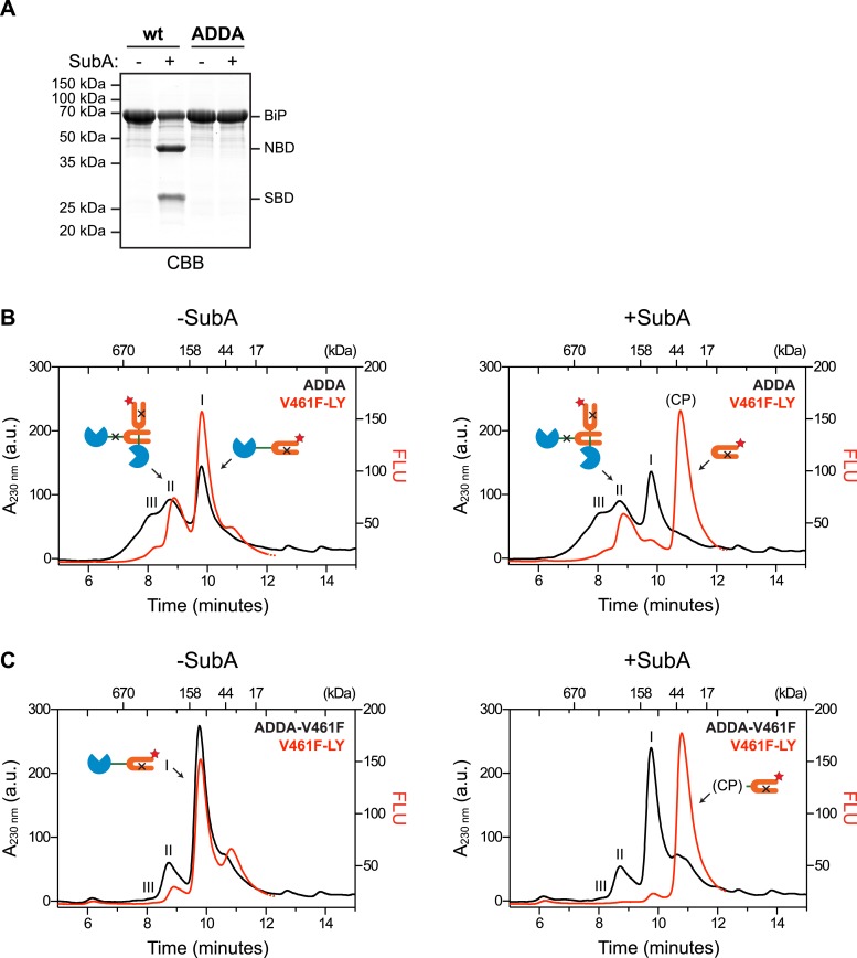
Figure 3. Cross-protomer engagement of the interdomain linker in BiP oligomers protects it against cleavage by SubA.

(A) Coomassie-stained SDS-gel of wildtype (wt) and ADDA mutant BiP (12 µM) before and after cleavage with SubA (0.25 ng/µl) for 15 min. Note that the ADDA mutation precludes cleavage by SubA. (B) Peptide bond absorbance trace (A230 nm, black) and fluorescence trace (in arbitrary fluorescence units, FLU) of lucifer yellow (LY) (Ex: 430 nm, Em: 525 nm; red) of purified ADDA mutant BiP (50 µM) incubated with tracer concentrations (1 µM) of lucifer yellow-labeled V461F mutant BiP (V461F-LY) and fractionated by size-exclusion chromatography (as in Figure 2) either before (-SubA) or after (+SubA) cleavage with SubA (36 ng/µl). Peaks I, II and III are marked as is the new fluorescent peak emanating from the V461F-LY cleavage product (CP). Note that fluorescent peak I, corresponding to the V461F mutant BiP monomer is attenuated after cleavage with SubA, whereas peak II, arising from hetero-oligomers of ADDA mutant BiP and V461F mutant BiP is relatively resistant. The peptide-bond absorbance trace (A230 nm), contributed largely by the ADDA mutant BiP, is unchanged by cleavage with SubA. (C) Experiment as in ‘B’ with purified double mutant ADDA-V461F BiP (50 µM) and lucifer yellow-labeled V461F mutant BiP (V461F-LY). Note the absence of a SubA-resistant fluorescent peak II in this sample.

DOI: http://dx.doi.org/10.7554/eLife.08961.008

Figure 3.

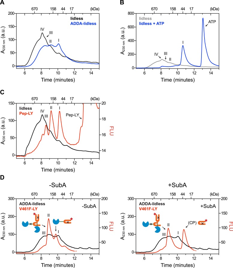
Figure 3—figure supplement 1. Removal of BiP's lid does not abrogate cross-protomer engagement of the interdomain linker in BiP oligomers.

Figure 3—figure supplement 1.

(A) Peptide bond absorbance traces (A230 nm) of purified BiP (50 µM) lacking its C-terminal ‘lid’ (∆554–654) without (lidless) or with a compounding ADDA mutation (ADDA-lidless) following fractionation by size-exclusion chromatography (SEC-3 column). The distinct peaks I, II and III (described in the legend to Figure 2) are indicated. (B) Trace as in ‘A’ of lidless BiP incubated with or without ATP (5 mM) before fractionation. Note the dissociation of oligomers upon exposure to ATP. (C) Peptide bond absorbance trace (A230 nm, black) and lucifer yellow (LY) fluorescence trace (Ex: 430 nm, Em: 525 nm; red) of purified lidless BiP (50 µM; as in ‘A’) supplemented with tracer concentrations of LY-labeled BiP-binding peptide (1 µM; as in Figure 2E) and fractionated as in ‘A’. The peak corresponding to the free peptide is indicated. Note the disproportionately diminished engagement of peptide by the larger lidless BiP oligomers, (D) Peptide bond absorbance trace of purified ADDA-lidless mutant BiP (50 µM) (A230 nm, black) and fluorescence trace (red) of tracer concentrations (1 µM) of fluorescent LY-labeled V461F mutant BiP (V461F-LY) fractionated by size-exclusion chromatography either before (-SubA) or after (+SubA) cleavage with SubA (36 ng/µl; as in Figure 3B). Peaks I, II and III are marked as is the new fluorescent peak emanating from the V461F-LY cleavage product (CP). Note that fluorescent peak I is attenuated upon treatment with SubA, whereas peak II is resistant. The peptide-bond absorbance trace, contributed largely by the ADDA mutant lidless BiP, is unchanged by SubA cleavage.