Figure 4.
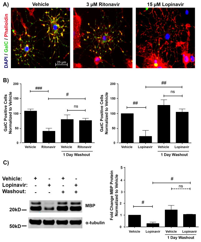
Antiretroviral inhibition of differentiation is reversible. (A) Oligodendrocyte precursor cells (OPCs) were treated with vehicle, 3 μM Ritonavir, or 15 μM Lopinavir at the time of differentiation. After 72 hours, the cells were fixed and stained for galactocerebroside (GalC, green) with DAPI (blue) and Phalloidin (red) for actin. Sample images reveal that cells in Ritonavir and Lopinavir treatments have elaborated morphology but lack the appropriate maturation marker GalC co-staining. Scale bar indicates 25 μm. (B) OPCs were treated as in A. After 72 hours, the washout group received new differentiation medium without antiretrovirals and was allowed to further mature for 24 hours. Cells were fixed and stained for GalC with DAPI. Cells were counted as in Figure 1 for 15 fields in 3 coverslips per condition at 40x magnification. Data are presented as mean ± SE, n = 4. One-way ANOVA followed by post-hoc Bonferroni correction compared vehicle and treatment conditions at the 2 time points (#p < 0.05; ##p < 0.01; ###p< 0.001, ns, not significant). (C) OPCs were treated with vehicle or 15 μM Lopinavir at the time of differentiation. After 72 hours, the washout group received new differentiation medium without treatment and was allowed to further mature for 24 hours. Cell lysates were immunoblotted for MBP protein. Representative Western blot image is shown. Quantification of band intensities normalized to a loading control reveals that Lopinavir-treated cells expressed less MBP protein compared with vehicle; however, this level rose to be comparable to vehicle 24 hours after washout. Data are presented as mean ± SE, n = 3. One-way ANOVA followed by post-hoc Bonferroni correction compared vehicle and treatment conditions at the 2 time points (#p < 0.05).

