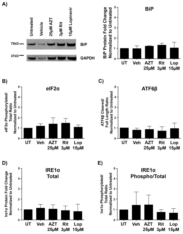
Figure 8.
The unfolded protein response is not triggered by antiretrovirals in developing oligodendrocyte precursor cells (OPCs). OPCs were treated with vehicle, 25 μM Zidovudine (AZT), 3 μM Ritonavir, or 15 μM Lopinavir at the time of differentiation. After 16 hours, cells were harvested for protein. Cell lysates were immunoblotted for protein alterations indicating activation of the unfolded protein response. (A) Representative Western blot images of BiP, with quantification of band intensities normalized to glyceraldehyde 3-phosphate dehydrogenase (GAPDH) loading controls from 3 independent biological replicates graphed. (B–E) Levels of phosphorylated compared to total eIF2a protein (B), cleaved compared to total ATF6β protein (C), total IRE1α protein (D), and phosphorylated compared to total IRE1α protein (E), were each analyzed from 3 separate biological replicates (n = 3). Quantification of band intensities normalized to GAPDH loading controls revealed no changes in any of these proteins following treatment with the antiretrovirals tested. Data are presented as mean ± SE. One-way ANOVA followed by post-hoc Dunnett’s multiple comparison test determined statistical significance.

