Figure 2.
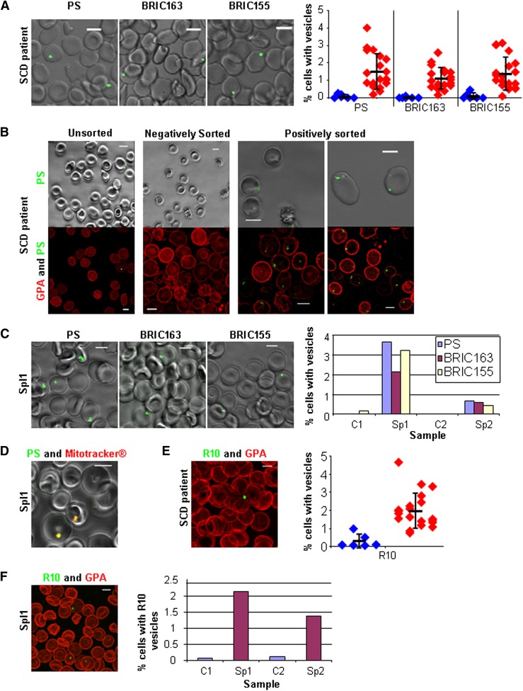
PS-exposed autophagic vesicles on red cells from peripheral blood in SCD and after splenectomy. (A) Live imaging of SCD red cells for PS, intracellular GPA (BRIC163), and intracellular AE1 (BRIC155); all green with quantitation from 20 SCD patients (red) and 8 controls (blue) imaging 5 random fields (average n = 439 per field), the thick horizontal line is the mean and standard deviation (SD) from the mean is shown. (B) Live imaging of SCD red cells after sorting for PS (green). Shown in phase overlay (upper panel) and as a 3D reconstruction (lower panel) dual-stained with GPA-546 (BRIC256) (red). (C) Live imaging of splenectomized patient 1 (Spl1, 3 years post-splenectomy) red cells for PS, intracellular GPA (BRIC163), and intracellular AE1 (BRIC155) (all green) with quantitation of Spl1 and splenectomized patient 2 (Spl2, 6 months post-splenectomy) imaging 5 random fields (average n = 375 per field). (D) Live imaging of red cells from Sp1 dual-stained with PS (green) and Mitotracker (red). (E) SCD red cells trypsin treated then fixed, permeabilized, and stained with R10 vesicles (green) and extracellular GPA (red) with quantitation from 20 SCD patients (red) and 8 controls (blue) imaging 5 random fields (average n = 381 per field), the thick horizontal line is the mean and SD from the mean is shown. (F) Spl1 red cells trypsin treated then fixed, permeabilized, and stained with R10 vesicles (green) and extracellular GPA (red) with quantitation from Spl1 and Spl2 with 2 controls imaging 5 random fields (average n = 280 per field). All scale bars are 5 µm.

