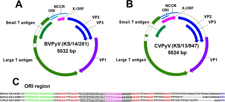
Fig 1. Schematic presentation of the genome organization of BVPyV (A) and CVPyV (B) and alignment of ORI region sequences of BVPyV strains (KS/14/281 and KS/13/999) and CVPyV strain KS/13/947 (C).
The positions of LTag introns are shown uncolored. Putative LTag binding sites (GAGGC pentanucleotides) are shown in red; AT-rich region/TATA box is shown in green; palindromic repeats shown in magenta; early palindrome (EP) region is underlined; LTag ATG codon is shown in blue (according [38, 39]).

