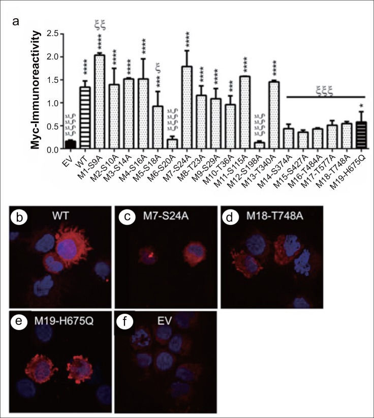
Fig. 2.
Effects of S/T→A mutations on AAH protein expression and subcellular localization: Huh7 cells were transiently transfected with wildtype (WT) or point-mutated (M#:S/T→A) N-Myc-AAH cDNA. Myc-empty vector (EV) served as a negative control. The M19-H675Q mutant, which disrupts AAH catalytic activity, served as a positive control. a Recombinant protein expression was measured by ELISA with the Myc antibody 24 h after transfection. The graph depicts mean (± S.E.M) levels of Myc. Inter-group comparisons were made by ANOVA with the post-hoc Fisher least significant difference (LSD) test. *p<0.05; **p<0.01; ***p<0.001; ****p<0.0001 for comparisons with EV, and ξp<0.05; ξξp<0.01; ξξξp<0.001; ξξξξp<0.0001 for comparisons with WT. (B-F) Representative results obtained by immunofluorescence staining and confocal imaging of cells transfected with (b) WT, (c) M7-S24A, (d) M18-T748A, (e) M19-H675Q, or (f) EV and stained by immunofluorescence with anti-Myc. Immunoreactivity was detected with biotinylated secondary antibody and streptavidin-conjugated DyLight 547 (red). Cells were counterstained with DAPI (blue). (Merged images: 600× magnification, 2× digital zoom).

