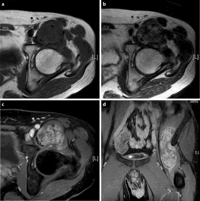
Fig. 1.
MRI showed a tumor extended from the medial aspect of the wing of the left ilium along the iliopsoas muscle to its site of insertion on the femur. The tumor was isointense on T1-weighted images (a) and heterogeneous hyperintense on T2-weighted images (b), and it was gadolinium enhanced (c, d).

