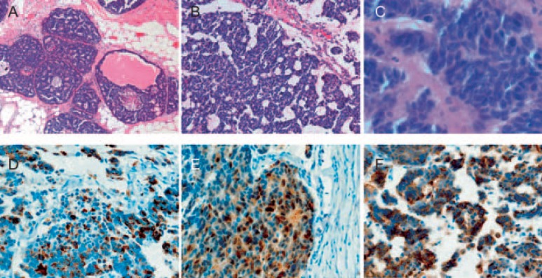
Fig. 2.
Pathological findings. The tumor is composed of solid nests and trabeculae without tubule formation. Large and polygonal shaped cells with faintly granular cytoplasm are separated by dense collagen palisading. The nuclear atypia of tumor cells is low. A, B low-power field, C high-power field (hematoxylin and eosin stain). D-F Tumor cells are positive for neuroendocrine markers. chromogranin A (D), neuron-specific enolase (E), and synaptophysin (F).

