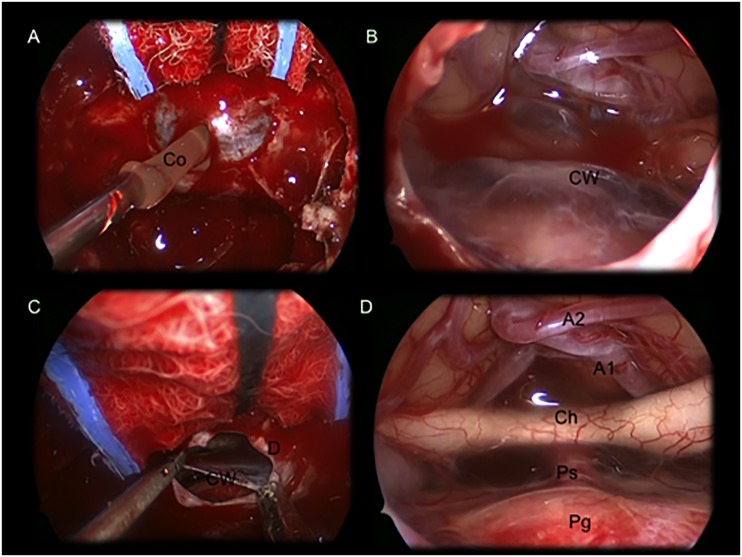
Fig 3. Intraoperative images showing a suprasellar Ratkhe’s Cleft Cyst removed via an extended endoscopic endonasalapproach.
(A) colloid suctioning after dural opening and exposure of the cyst’s wall (B)Imagine showing the cyst’s wall covering the neurovascular structures of the suprasellar area. (C) cyst wall removal with a forceps and aspirator. (D) after cyst wall removal it is possible to identify: A1 and A2; optic chiasm with optic nerves; pituitary stalk and gland. Co: colloid; CW: cystwall; D: dura mater; Ch: optic chiasm; Ps: pituitary stalk; Pg: pituitary gland; A1: A1 segment of the anterior cerebral artery; A2: A2 segment of the anterior cerebral artery.

