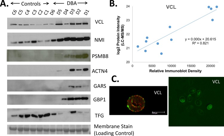
Fig 7. Validation of proteomics data using western blot and immunofluorescence.
A) Immunoblot of a number of proteins that were significantly changed in Hb-depleted erythrocyte cytoplasm between DBA patients and healthy donors. Equal protein loading and consistent electrotransfer of samples for the western blots were confirmed by staining the entire PVDF membrane using MemCode, a reversible total protein stain, immediately prior to performing the western blot as shown by the representative 30 kDa protein visualized by membrane stain in the bottom panel. Images for MemCode staining of all other immunoblots are shown in S2B Fig) Correlation between VCL intensity values from LC-MS/MS label-free quantitation and relative density from immunoblot analysis. C) Immunofluorescence of VCL and Band 3 within erythrocytes from patient D3 visualized with confocal microscopy (left, Band 3 in red and VCL in green) and fluorescence microscopy (right, VCL in green).

