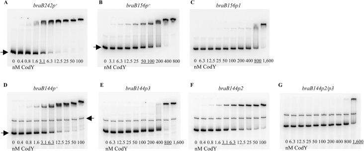
Fig 4. Gel shift assays of CodY binding to braB fragments.
The braB242p + (A), braB156p + (B), braB156p1 (C), braB144p + (D), braB144p3 (E), braB144p2 (F), and braB144p2/3 (G) DNA fragments labeled on the template strand were incubated with increasing amounts of purified CodY in the presence of 10 mM ILV. The fragments were obtained by PCR with oligonucleotides oBB67 and oBB102 (A), oBB358 and oBB102 (B, C), and oBB422 and oBB102 (D-G) and their positions in the gel are indicated by right-pointing arrows. The unspecific DNA fragment, present in some panels, is indicated by a left-pointing arrow. CodY concentrations used (nM of monomer) are reported below each lane; concentrations corresponding to the apparent KD for binding are underlined.

