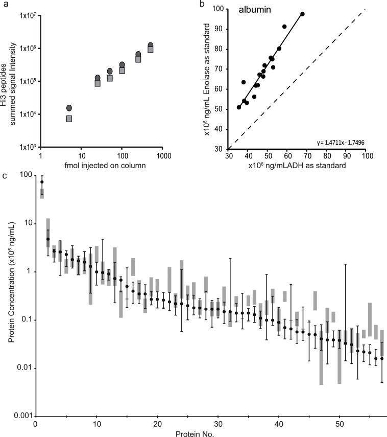
Fig 1. HI3 peptide quantitation with a single protein digest standard and digest standard comparison.
(a) Summed signal intensity of the protein digest standard ENO1 (grey square) and ADH1 (dark grey circle) added at increasing concentrations to a plasma digest. (b) Quantitation of albumin using either ENO1 or ADH1 as the internal standard in 17 indivual samples. The regression line (solid black) and its formula, obtained by ordinary least squares linear regression, is depicted, with the dashed line representing perfect correlation. (c) 57 proteins from Table 2 for which reference ranges from literature were available, are ordered according to their median concentration determined by HI3 peptide quantitation (dark grey squares, quantified in ≥ 11 out of 31 samples). Error bars indicate the minimal and maximum value measured in the plasma samples. The reference ranges (grey boxes) are taken from Hortin et al. [42]. Protein no. correspond to the numbers given in Table 2.

