
Fig. 3.
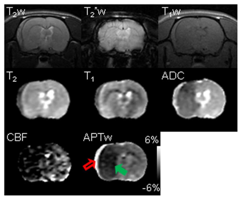
Multi-parametric MRI features of subarachnoid hemorrhage (red open arrow) and cerebral ischemia (green solid arrow) in a rat at one hour post MCAO. Subarachnoid hemorrhage was induced by injury to the vessel when preparing the MCAO model. This rat was sacrificed after MRI, and the subarachnoid hemorrhage was confirmed by the direct observation. The display windows are T2 (25 to 75 ms), T1 (1 to 2 sec), ADC (0.4 to 1.2×10⁻⁹ m2/sec), blood flow (0 to 120 ml/100g/min), and APTw (-6% to 6% of the bulk water signal intensity). APTw MRI demonstrated the starkest contrast between the hematoma and ischemia (hyperintense or hypointense with respect to the contralateral tissue, respectively).