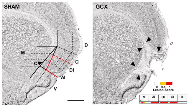
Figure 12.

Example of the lesion analysis system applied to the study of the functional consequences of gustatory cortex lesions. Left: a Nissl-stained cross section of an intact rat brain at the level of +1.2 mm relative to bregma. A grid is placed over the region of interest and an experimenter blinded to the behavioral outcome of the animal scores each grid cell for the presence of a complete (1.0), partial, (0.5), or no lesion (0). Abbreviations: AI, agranular insular cortex (dorsal to the rhinal fissure); C, claustrum (outlined column just medial to AI, GI, and DI); D, dorsal to insular cortex; DI, dysgranular insular cortex; GI, granular insular cortex; M, medial to claustrum; V, ventral to rhinal fissure; (Scale bar: 1.0 mm.). Right: an example of how a lesion would be scored. Reprinted from Schier LA, Hashimoto K, Bales MB, Blonde GD, Spector AC. (2014) High-resolution lesion-mapping strategy links a hot spot in rat insular cortex with impaired expression of taste aversion learning. Proceedings of the National Academy of Sciences of the United States of America. 111: 1162–1167 with permission.
