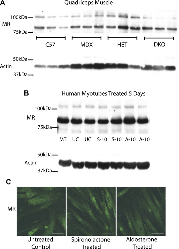
Figure 3.
MR protein levels are maintained in dystrophic muscle and agonist vs. antagonist treated human cells. A) Representative Western blots of quadriceps muscles from 3 biologic replicates are shown comparing MR protein levels from equivalent amounts of protein homogenates from C57BL/10 wild-type (C57) mice, dystrophin-deficient mdx (MDX) mice, het (HET) mice, and dystrophin/utrophin-deficient double knockout (DKO) mice. B) MR was also detected by Western blots of human primary muscle (HSMM) myotubes differentiated for 5 d (MT) and then treated for 5 d with 10 µM of MR antagonist spironolactone (S-10), MR agonist aldosterone (A-10), or ethanol vehicle untreated control (UC). For (A) and (B), 35 µg of total protein was used for each sample. Western blots used combination of MR-specific monoclonal antibodies MR1-18 1D5 or MR1-18 6G1 and MRN 2B7 (31) (full-length MR predicted molecular weight, ∼107 kDa) or α-sarcomeric actin antibody (loading control; predicted molecular weight, ∼42 kDa). C) MR localization is similar after treatment with MR antagonist and agonist. MR was detected by immunofluorescence staining (green) on human primary (HSMM) myotubes differentiated for 5 d then treated for 48 h with 10 µM of MR antagonist spironolactone or MR agonist aldosterone. Staining of subset of nuclei was present in all 3 groups. MR staining is similar with longer 5 d treatments (data not shown). Scale bars, 50 µm.

