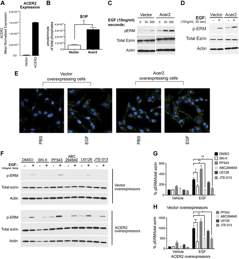
Figure 5.
Increased intracellular S1P production is sufficient to promote ERM activation. A) mRNA of HeLa cells overexpressing empty vector or ACER2 was assessed by quantitative RT-PCR. B) Cellular lipids were directly extracted in organic solvents from HeLa cells overexpressing empty vector or ACER2. S1P levels were analyzed by tandem liquid chromatography/MS. C) HeLa-ACER2-TET-ON and HeLa-vector control cells were treated with tetracycline (5 ng/ml) for 12 h. These cells were then starved for 4 h prior to treatment with EGF (10 ng/ml) for either 30 s or 5 min. pERM levels were then assessed by immunoblotting (C) and by confocal microscopy (E). The green color corresponds to pERM levels labeled with Alexa 488 fluorophore antibody, and the blue color corresponds to DAPI staining the nuclei (E). D) HeLa cells were transiently transfected with either vector control or ACER2 overexpressing DNA for 24 h. Cells were then starved for 4 h prior to treatment with EGF (10 ng/ml) for 5 min. pERM levels were then assessed by immunoblotting. Total ezrin and actin were also included as loading controls. F) HeLa cells stably overexpressing vector or ACER2 were starved overnight. These cells were then pretreated with Ski-II (1 μM), PF543 (100 nM), ABC294640 (10 μM), U0126 (10 μM), or JTE-013 (5 μM) prior to stimulation with EGF (10 ng/ml) for 5 min. G and H) Quantification of the ratio of pERM:total ezrin in (F) was performed using ImageJ software. The data represent means ± se of 3 independent experiments. *P < 0.05; **P < 0.01.

