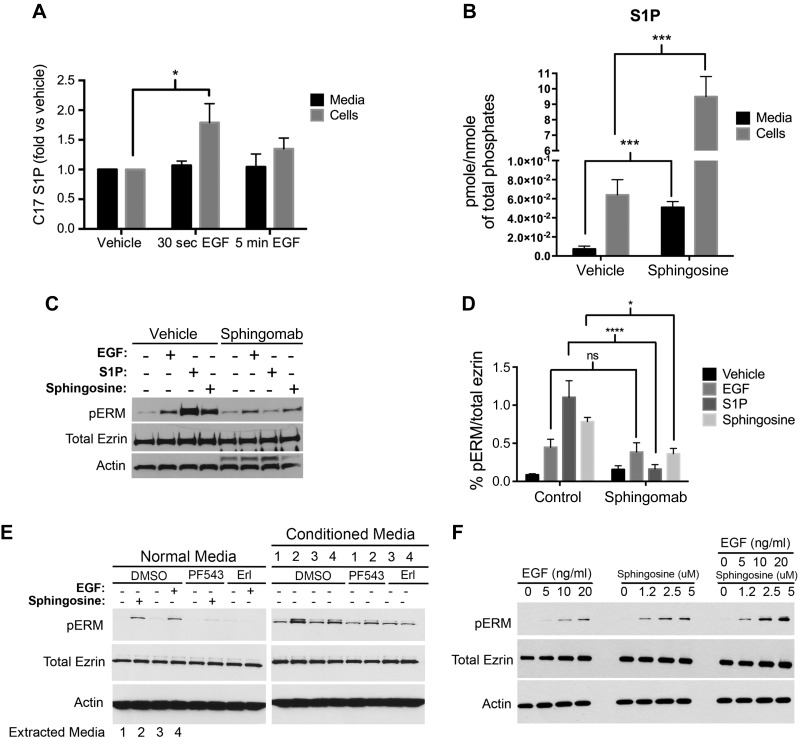
Figure 6.
ERM phosphorylation does not require extracellular S1P production. A) HeLa cells were prelabeled for 30 min with 250 nM C17-Sph. EGF (10 ng/ml) was then added for 30 s or 5 min. C17-S1P levels from medium and cells were then analyzed via mass spectroscopy. B) HeLa cells were starved overnight, then treated with 5 μM sphingosine for 5 min. Endogenous S1P levels from medium and cells were then analyzed via mass spectroscopy. The data represent means ± se of 3 independent experiments performed in duplicates. C) HeLa cells were pretreated for 1 h with either PBS or 50 μg/well Sphingomab prior to treatment with EGF (10 ng/ml), or sphingosine (5 μM), or S1P (100 nM) for 5 min. pERM levels were then assessed by immunoblotting. Total ezrin and actin were also included as loading controls. D) Quantification of the ratio of pERM:total ezrin in (C) was performed using ImageJ software. The data represent means ± se of 2 independent experiments. E) HeLa cells were pretreated with DMSO, PF543 (100 nM), or erlotinib (Erl; 100 nM) for 1 h, then followed by treatment with either sphingosine (5 μM) or EGF (10 ng/ml) for 2 min. Media from DMSO-pretreated cells (with EGF, Sph, or vehicle treatment), named conditioned media and labeled 1–4, were then added on top of HeLa cells that are pretreated with DMSO, PF543 (100 nM), or erlotinib (100 nM). The reasoning lies in that PF543 treatment will inhibit sphingosine conversion to S1P and that erlotinib will inhibit EGF effect; thus, any effect seen after the addition of the conditioned media will be the result of S1P presence in these media. pERM levels were then assessed by immunoblotting as a marker of S1P presence. Total ezrin and actin were also included as loading controls. F) HeLa cells were plated for 24 h and then starved for 4 h prior to treatment with increasing doses of sphingosine, EGF, or both. pERM levels were then assessed by Western blotting. Total ezrin and actin were also included as loading controls. ns, not significant; *P < 0.05; ***P < 0.001; ****P < 0.0001.

