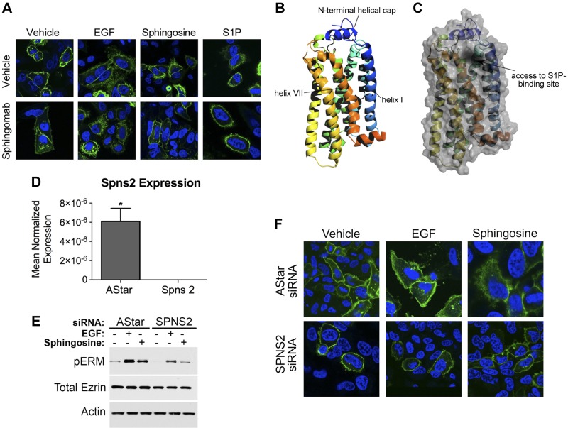
Figure 7.
Spns2 is partially required for EGF-mediated ERM phosphorylation and S1PR2 internalization. A) HeLa cells were transfected with S1PR2-GFP for 24 h. Following transfection, cells were starved for 4 h and then pretreated for 1 h with PBS or Sphingomab. Cells were treated with 100 nM S1P, 10 nM EGF, or 5 μM sphingosine. Cells were then fixed, and nuclei were stained with DAPI (blue). Images were taken using the Leica SP8 confocal microscope. B and C) Structure model for S1PR2 generated using the I-TASSER server (B). Cartoon with surface overlay, demonstrating the proposed point of access for S1P from the membrane into its receptor-binding site, is shown in (C). D) HeLa cells were treated with 20 nM AStar or Spns2 siRNA for 48 h, and Spns2 mRNA was then assessed by quantitative RT-PCR. *P < 0.05. E) HeLa cells were treated with AStar or Spns2 siRNA for 48 h. Cells were then starved for 4 h prior to treatment with EGF (10 ng/ml) or sphingosine (5 μM) for 5 min. pERM levels were then assessed by immunoblotting. Total ezrin and actin were also included as loading controls. F) HeLa cells were treated with AStar or Spns2 siRNA for 48 h. HeLa cells were then transfected with S1PR2-GFP, 24 h following siRNA transfection. Cells were starved for 4 h and then treated with 10 nM EGF, or 5 μM sphingosine. Cells were then fixed, and nuclei were stained with DAPI (blue). Images were taken using a Leica SP8 confocal microscope.

