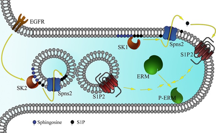
Figure 8.
Schematic representation of the proposed model for EGF-induced ERM phosphorylation. EGF treatment activates SK2 localized on the cytosolic side of intracellular vesicles. This will lead to S1P being produced and transported by Spns2 (as well as other ABC transporters) to the inner side of these vesicles. These vesicles will then fuse with S1PR2-containing vesicles or with the plasma membrane. This is followed by S1PR2 activation and ERM phosphorylation, and filopodia formation as shown in this cellular extrusion. On the other hand, sphingosine treatment causes S1P production by SK1 localized on the plasma membrane. S1P will then be exported to the extracellular milieu by Spns2, leading to S1PR2 activation and ERM phosphorylation.

