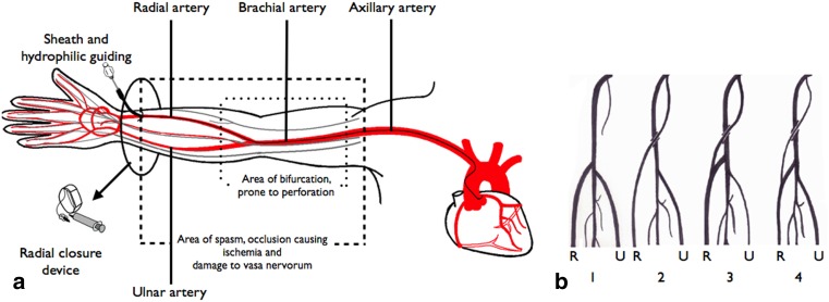
Fig. 1.
The anatomy of the upper extremity (a) and its variations (b). a The anatomy of the arteries (red line) and nerves (grey line) of the arm leading to the heart. The area where the bifurcation of the radial artery might occur is accentuated; this area is prone to perforation (inner dashed box). The area where spasm, occlusion or damage to vasa nervorum occurs is also highlighted (outer dashed box). Hydrophilic guiding catheters and special radial access closure devices might reduce the incidence of these complications and could diminish the impact on upper extremity function. b Frequent variations of the take-off of the radial artery. The radial artery ® and ulnar artery (U) are illustrated. 1. Radial artery arising from the brachial artery. 2. Independent radial artery arising from the axillary artery. 3. Radial artery arising from the axillary artery with a contribution from the brachial artery. 4. Slender artery arising from the axillary artery continuing as the radial artery. The major blood supply to the radial artery is supplied by the brachial artery. This type is highly susceptible to perforation.

