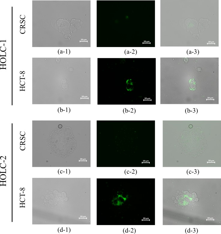
FIG. 6.
Microscopic images of FAM-labeled HOLC-1 and HOLC-2 peptides bound to CRSCs and HCT-8 cells. (a-1) and (c-1) Bright-field images of CRSCs. (a-2) and (c-2) Fluorescent microscope images of HOLC-1- and HOLC-2-bound CRSCs, respectively. (a-3) and (c-3) Merged images of HOLC-1- and HOLC-2-bound CRSCs, respectively. (b-1) and (d-1) Bright-field images of HCT-8 cells. (b-2) and (d-2) Fluorescent microscope images of HOLC-1- and HOLC-2-bound HCT cells, respectively. (b-3) and (d-3) Merged images of HOLC-1- and HOLC-2-bound HCT-8 cells.

