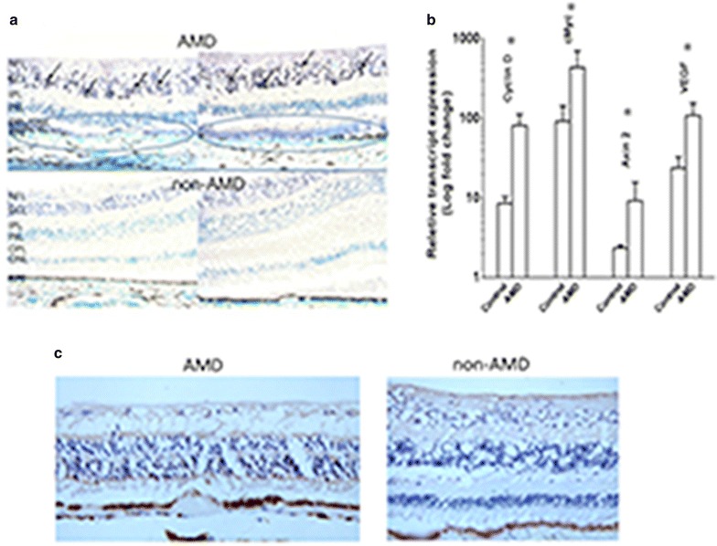
Fig. 1.

Wnt signaling molecules in human retina. a Immunochemistry of phosphorylated LRP6 found higher activated LRP6 in the ganglion cell layer (brown-blackish staining, arrows) of the retinal sections of AMD maculae (upper panel) than in non-AMD macular tissues (bottom panel). Atrophic/degenerative photoreceptors and abnormal RPE cells are also noted in the macula of AMD specimens (blue circles). These areas also illustrate stronger immunoreactivities compared to the normal macula in the lower panel. NFL nerve fiber layer, GCL ganglion cell layer, IPL inner plexiform layer, INL inner nuclear layer, OPL outer plexiform layer, ONL outer nuclear layer. b mRNA expression of microdissected macular cells from human retinal section was measured by RT-PCR. Higher expression of CYCLIN D, cMYC, AXIN2, and VEGF was observed in AMD patients compared to non-AMD controls (N = 5, Error bar mean ± SEM); *p < 0.05. c Immunochmistry of β-catenin in the macula. Higher expression of β-catenin was illustrated in AMD macular region compared to non-AMD macular area
