Fig. 4.

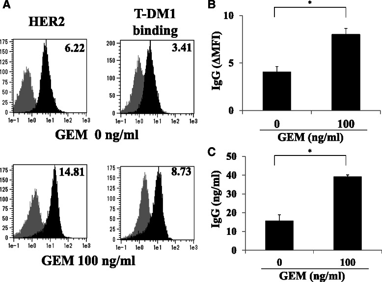
GEM treatment increased HER2 expression and resulted in increased T-DM1 binding to MIA PaCa-2 cells. a MIA PaCa-2 cells were either untreated or treated with GEM (100 ng/ml) for 2 h, washed with PBS and incubated in fresh medium for 48 h. The cells were detached by trypsin-EDTA treatment, untreated or treated with T-DM1 (30 μg/ml) for 1 h and then analyzed by flow cytometry using PE-labeled anti-human IgG. Left: HER2 expression in untreated or GEM-treated MIA PaCa-2 cells. Right: T-DM1 binding to untreated or GEM-treated MIA PaCa-2 cells. The grey and black areas indicate the isotype control and the specific antibody, respectively. The values on the left indicate ΔMFI for HER2, representing the MFI for HER2 minus that of the isotype control. The values on the right indicate ΔMFI for human IgG, representing the MFI for PE-labeled anti-human IgG minus that of the isotype control. b Untreated or GEM-treated MIA PaCa-2 cells were incubated with T-DM1 as described in (a), and T-DM1 binding was examined by flow cytometry using PE-labeled anti-human IgG. The ΔMFI of human IgG was calculated as the MFI of PE-labeled anti-human IgG minus that of the isotype control. *P < 0.01 (n = 3). c Untreated or GEM-treated MIA PaCa-2 cells were incubated with T-DM1 as described in (a). The cells (1x10⁶) were lysed, and the amount of human IgG binding to the cells was determined by ELISA. *P < 0.01 (n = 3)
