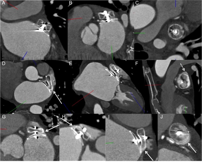
Figure 4.
Contrast-enhanced CT images: (A–C) Amulet case with good alignment of lobe perpendicular to the neck of the LAA, with complete occlusion of the LAA. (D–F) ACP case with off-axis lobe (not perpendicular to the LAA neck) (white arrow in D) and superior gap (dashed arrow in F) causing residual contrast patency of LAA. (G) Example of ACP case with off-axis on the right side (arrow) with contrast leak through that edge, whereas there was no lobe leak on the left side. (H) Example of ACP case with off-axis lobe and contrast leak through the superior edge (arrow) (bottom half of lobe with no contrast opacification). (I) ACP lobe off-axis inferiorly (arrow) with contrast leaking through the edge and gap (arrow in J).

