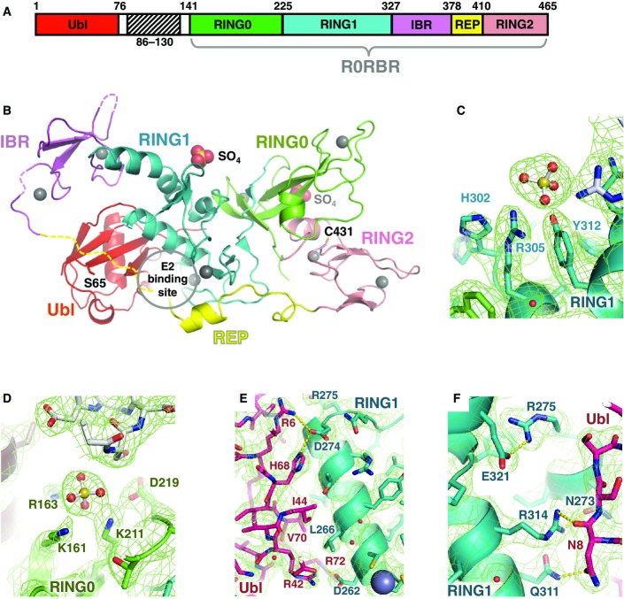
Figure 1.
- A Domain organization of Parkin, showing location of Δ86–130 deletion and definition of R0RBR module.
- B Structure of Δ86–130 Parkin. The sites of phosphorylation on Ser65 of the Ubl domain and the catalytic residue Cys431 are indicated along with the bound sulfates.
- C, D Polar contacts and residues around the bound sulfate ions. The sulfate in the first site has B-factor of 40 Å2 and the second has a B-factor of 80 Å2.
- E, F Close-up views of the interaction between the Ubl (red) and RING1 domains (cyan).

