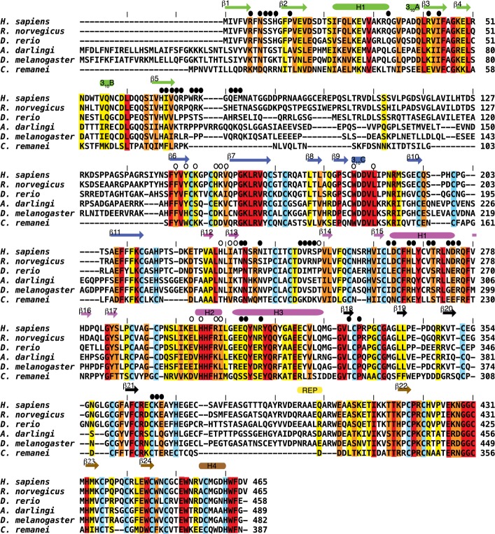
Figure EV1. Structure-based sequence alignment of parkin from different species: human, rat, zebrafish, mosquito, fruit fly and nematode.
Secondary structure elements are indicated above the alignment and coloured according to domains where the Ubl is in green, RING0 in blue, RING1 in magenta, IBR in black, REP element in yellow and the RING2 in brown. Zinc-coordinating residues are shaded in cyan. Conserved residues are shaded in red, conservative substitutions in orange and semi-conserved residues in yellow. Closed circles denote residues involved in the Ubl interface, and open circles denote those in the RING0/RING1 interface.

