
Figure 8.
Notch-activated antral polyps exhibit increased stem and progenitor cell marker expression
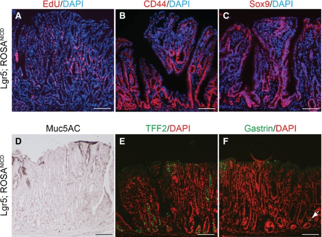
- A–C Paraffin sections from Lgr5; ROSANICD stomach 6–12 months post-TX were stained for (A) proliferation by EdU incorporation (red) or immunostained for (B) CD44 or (C) Sox9. DAPI (blue) was used as a nuclear counterstain. Scale bars: 100 μm.
- D–F Immunohistochemical analysis of (D) surface mucous cells (Muc5ac), (E) deep mucous cells (TFF2) and (F) endocrine cells (Gastrin) in Lgr5; ROSANICD mice 6–12 months post-TX. Compare Fig4 for normal tissue staining. Scale bars: 100 μm.