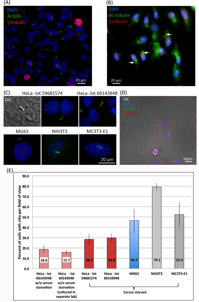
Figure 1. Primary cilia are frequent on HeLa cancer cells.
(A, C) Two different lots of HeLa cells purchased from ATCC stained positive for Arl13b (green), a protein localized with high specificity to the axonemes of cilia. At the base of the cilia, centrioles visualized using an antibody targeting γ-tubulin (red) are clearly detectable as well. Cell nuclei are stained with DAPI (blue). (B) A more traditional stain for primary cilia, acetylated-tubulin (green) and γ-tubulin (red), also stains primary cilia (arrows) but not as distinctly as with antibodies directed against Arl13b. (C) Primary cilia were also detected using DIC. NIH3T3 cells, reported as ‘the gold standard’ for primary ciliated cells, and MC3T3-E1 cells both exhibited a similar staining pattern to HeLa cells. (D) Fluorescent Arl13b (green), γ–tubulin (red), and DAPI (blue) stains merged with a DIC image demonstrate that even at very low density HeLa cells form primary cilia. (E) Quantitative analysis of primary cilia detected in different lots of starved and un-starved HeLa cells, as well as in starved MG63 osteosarcoma, NIH3T3 fibroblasts, and MC3T3-E1 pre-osteoblast cells. The number of HeLa cells that stain positive for primary cilia that we detected is significantly higher (28.6% after starvation, 18.6% in un-starved cells) than had been reported previously (<2%). In NIH3T3 and MC3T3-E1 comparable values to previously published reports were detected (Alieva et al., 1999; Malone et al., 2007; Schneider et al., 2005).

