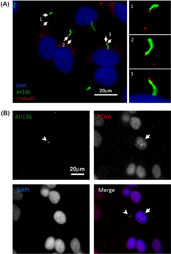
Figure 2. Actively proliferating HeLa cells also have primary cilia.
(A) Arl13b (green) and γ-tubulin (red) staining reveals several HeLa cells with two centrioles (depicted with arrows) indicating that these cells are in either the S- or G2- phase of the cell cycle. Higher magnifications of the regions containing the cilium and centrioles are to the right. (B) Arl13b staining (depicted with arrowhead) together with PCNA staining further indicates that proliferating cells have primary cilia as indicated by the distinctive punctate PCNA S-phase staining pattern in the nuclei of cells (depicted with arrow) containing primary cilia. Cell nuclei are stained with DAPI (merged panel: Arl13b, green; PCNA, red; nuclei, blue).

