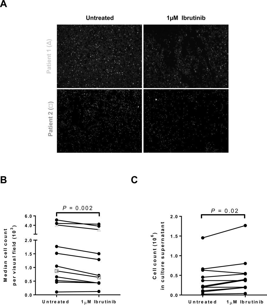
Figure 3.
Release of adhered cells by ibrutinib in vitro. A, Microphotographs show results from two representative patients. CLL PBMCs were adhered to fibronectin coated plates that were left untreated or incubated for 1 hour with ibrutinib. B, The median cell count per visual field on untreated and ibrutinib treated plates is shown. Lines connect results obtained with cells from the same patient (n=11). Symbols correspond to the representative patients depicted in panel A. C, The number of CLL PBMCs released from the fibronectin coated plate into the media after 1 hour of incubation with ibrutinib compared to control is shown. Lines connect results obtained with cells from the same patient (n=11). All Statistics were determined by a Wilcoxon matched pairs test.

