Figure 5.
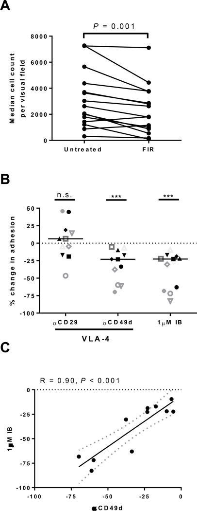
The role of BTK and VLA-4 in CLL cell adhesion to fibronectin. A, The median count of cells adhering to fibronectin coated plates per visual field is shown pre-treatment (Pre) and after 1 hour of incubation with 500nM firategrast (FIR; VLA-4 antagonist). Lines connect results obtained with cells from the same patient (n=15), P value by Wilcoxon matched pairs test. B, The percent change in cell adhesion normalized to untreated control is shown for samples (n=11) treated for 1 hour with blocking antibodies against either CD29 (αCD29) or CD49d (αCD49d) the two components of VLA-4, or with 1µM ibrutinib (IB). Each symbol represents a different patient. Lines represent median. P value determined by a Wilcoxon matched pairs test: ***P<0.001. C, Pearson’s correlation of the reduction in cell adhesion by ibrutinib (IB) and αCD49d. Each circle represents a different patient (n=11); dotted lines represent 95% confidence interval.

