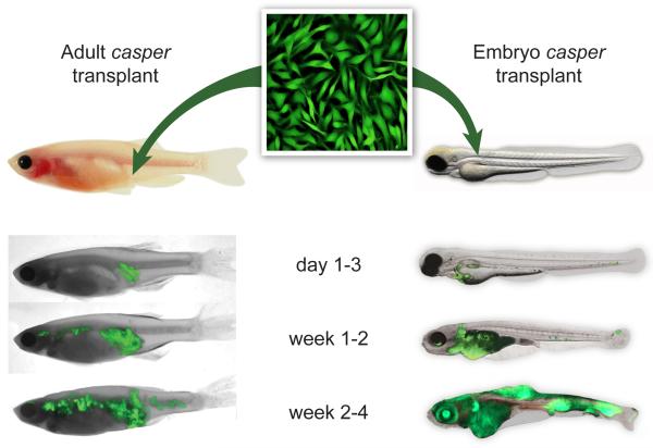
Figure 2.
Evaluating metastasis of the ZMEL1 line using transplantation into the transparent casper recipient line. ZMEL1-GFP cells can be transplanted either subcutaneously into the flank of an irradiated casper recipient (left) or directly into the vasculature of an unirradiated casper embryo at 2 days post fertilization (right). The fish are then imaged over a period of ~1 month using GFP and brightfield imaging. Representative fish for both assays are shown. For the adults, ~500,000 cells were transplanted, and for the first 1-3 days after transplant the cells remain localized, but by weeks 1 to 4, they widely disseminate anteriorly and posteriorly from the initial implant site. A similar pattern is seen in the embryo transplants, but because the cells are injected directly into the circulation, extravasation and formation of disseminated masses occurs more rapidly.

