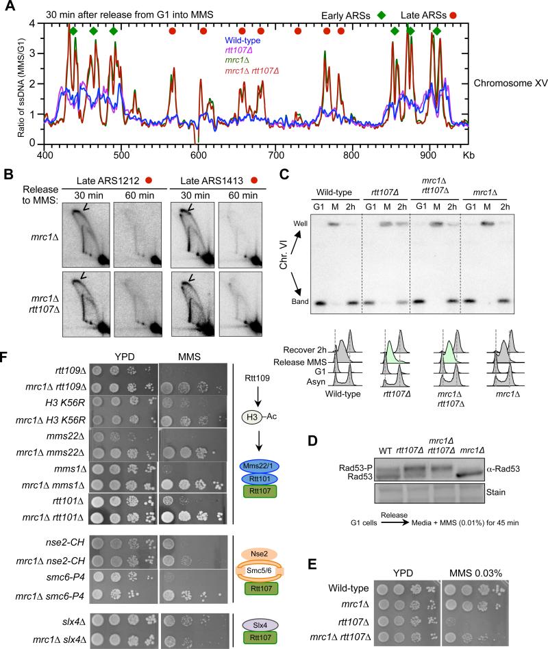
Figure 5. mrc1Δ increases global late origin firing and rescues defects due to the loss of Rtt107 and its interacting E3s.
(A) Replication fork-associated ssDNA profiles show that mrc1Δ increases late origin firing. The profiles of a segment of Chromosome XV are shown as representative images of the genome-wide data. G1 cells were released into media containing MMS for 30 min. Relative amount of ssDNA was determined as the ratio of fluorescent signal from the 30 min S phase sample to that from the G1 sample. The early and late origins are indicated by green diamonds and red dots, respectively.
(B) 2D gel results verify that mrc1Δ leads to firing at two late origins, ARS1212 and ARS1413, in MMS. Experimental procedures and labels are identical to those in Figure 4.
(C) mrc1Δ increases chromosomal replication in rtt107Δ cells. G1 cells were released into media containing 0.03% MMS for 1 h (Release MMS; M), then recovered in normal media for 2 h (Recover 2 h; 2h). FACS shows the bulk DNA synthesis of each strain, with mrc1Δ rtt107Δ exhibiting faster progression than rtt107Δ cells (green). Chromosomes separated by PFGE were examined by Southern blot using a probe specific to Chr. VI. Quantification of the results seen Figure. S5B.
(D) mrc1Δ does not reduce Rad53 phosphorylation levels in rtt107Δ cells. Experiments were done as in Figure 1E.
(E-F) mrc1Δ suppresses the MMS sensitivity of cells defective in Rtt107 or its interacting E3s, but not Slx4. MMS sensitivities were examined as in Figure 2B. (E) Diagrams on the right summarize the relevant complexes and proteins examined. MMS concentrations for panels containing the following mutants are rtt109Δ: 0.02%; H3 K56R and mms22Δ: 0.01%; rtt101Δ, mms1Δ and nse2-CH: 0.03%; smc6-P4: 0.003%, and slx4Δ: 0.05%.
See also Figure S5.

