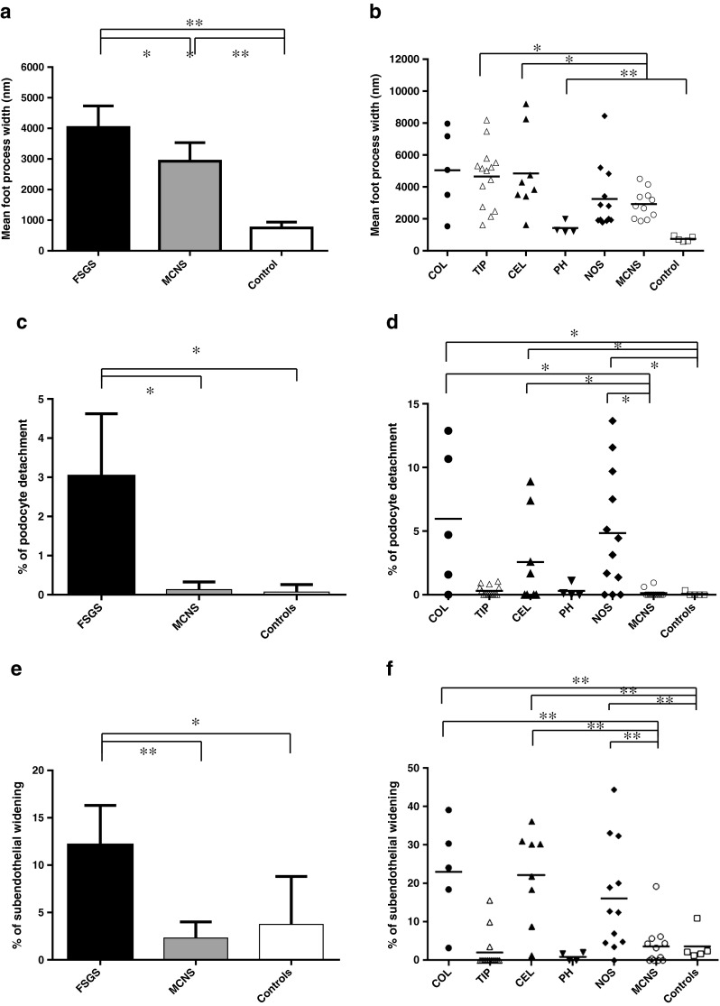
Fig. 3.

a Mean foot process width (a, b), percent podocyte detachment (c), and percent subendothelial widening (e), with the 95 % confidence interval in electron micrographs of the kidneys from patients with focal segmental glomerulosclerosis (FSGS), patients with minimal change nephrotic syndrome (MCNS), and control donors. b Mean foot process width, percent of GBM length with PD (% of podocyte detachment; d) and percent of GBM length with SW (% of subendothelial widening; f) in electron micrographs of the kidneys from patients with each variant of FSGS, patients with MCNS and control donors. *P < 0.05; **P < 0.01. COL collapsing variant, TIP tip variant, CEL cellular variant, PH perihilar variant, NOS not otherwise specified variant
