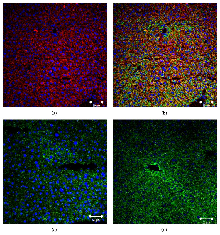
Figure 4.
Immunofluorescent analysis of SR-BI localization in liver of normal, Tie2-Scarb1 × Scarb1-KO, and Scarb1-KO mice. Liver sections from normal (a, b) and Tie2-Scarb1 × Scarb1-KO mice (c) were stained for SR-BI (red) and cytokeratin 8–18 (green). Merge image for normal mouse (b) demonstrates strong presence of SR-BI in hepatocytes (b, yellow signal) and absence of detectable level of SR-BI protein in liver of Tie2-Scarb1 × Scarb1-KO mice (c). In Scarb1-KO mice there was no detectable level of SR-BI protein (d, staining for SR-BI). Blue = DAPI.

