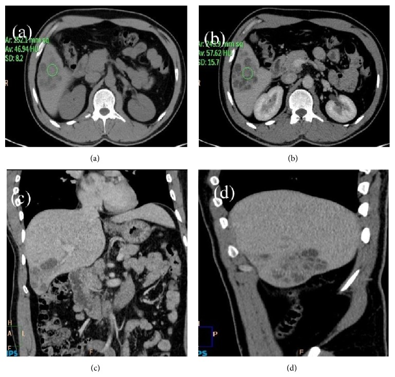
Figure 1.
(a) Plain CT image of abdomen: axial view showing ill-defined, mixed density mass lesions in segment V of liver. Hounsfield unit at noncystic part is 47 in the center of the lesion. (b) Postcontrast CT image of abdomen: axial view showing ill-defined heterogeneously enhancing conglomerated mass lesions in segment V of liver. Hounsfield unit at noncystic part is 57 in the center of the lesion. (c) Postcontrast CT image of abdomen: coronal view showing ill-defined conglomerate mass lesions in segment V of liver. Remainder of abdomen and visualized part of lungs are devoid of any significant pathology. (d) Postcontrast CT image of abdomen: sagittal view showing ill-defined conglomerated lesions in segment V of liver. There is mild enlargement of the liver.

