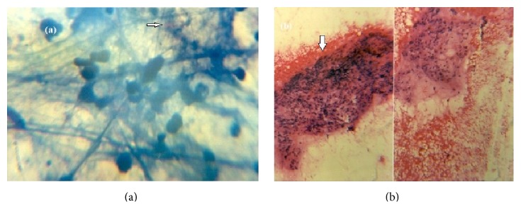
Figure 2.

(a) Immunofluorescence using AFB (ICC, ∗100x) confirms that the antibody specifically stains one or a few mycobacteria (white arrow) inside the cytoplasm of cells. (b) On histopathological analysis (H and E, ∗100x) there was collection of ill-defined epithelioid cells, lymphoid cells, and multinucleate giant cells with central area of necrosis within (white arrow).
