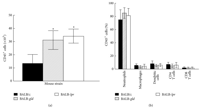
Figure 5.
Inflammatory infiltrate in the corneas of BALB-gld and BALB-lpr mice displays significant increased CD45+ cells, though there are no qualitative differences in subpopulations of CD45+ cells. Mice were infected in one eye with HSV-1 and reactivated 8 weeks later by UV-B irradiation. The HSV-infected corneas were removed at days 17 and 23 after irradiation from mice with severe HSK disease and disaggregated into single-cell suspensions and stained with anti-CD45 (a). The CD45+ cells were gated and further analyzed for staining with anti-CD4, CD8α, Gr-1, CD11b, CD11c, and F4/80 mAb (b). Cells were analyzed by flow cytometry. Data represents 4 to 6 corneas per group. Significant differences were seen in CD45+ cells (a) (P < 0.05–0.02), but not for the percentages of CD45+ subsets (b) (P > 0.05).

