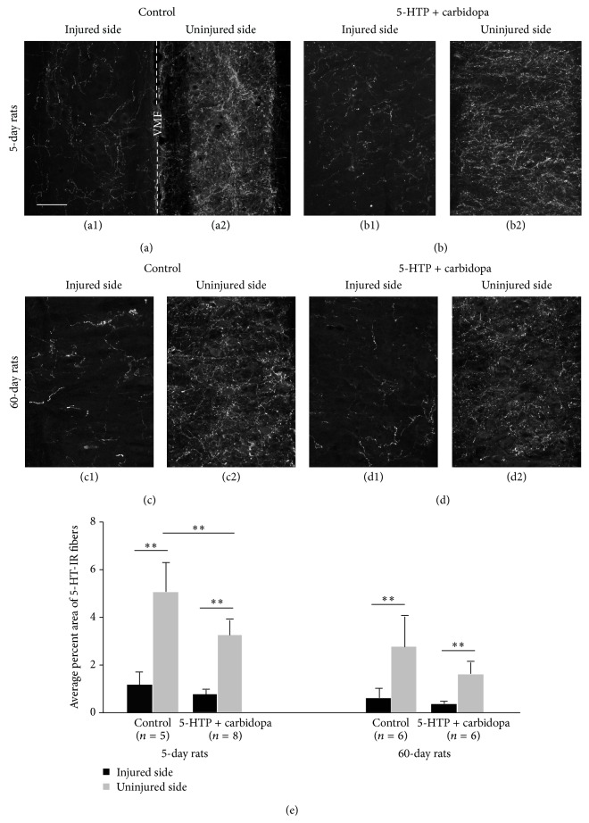
Figure 2.
5-HT-immunoreactive (IR) nerve fibers in the ventral horn motoneuron region in the sacrocaudal spinal cord on the injured and uninjured side in different animal groups. (a1–d2) Photomicrographs of 5-HT-IR fibers in 5-day untreated group (a1, a2) and 5-HTP plus carbidopa treated group (b1, b2) and in 60-day untreated group (c1, c2) and 5-HTP plus carbidopa treated group (d1, d2). (a1) and (a2) were from the same horizontal section. Dashed line indicates the midline. VMF: ventral median fissure. Other pairs of photomicrographs were from different sections. Scale bar in (a1), 100 μm. (e) Quantitative data of the density of 5-HT-IR fibers in different animals groups.∗∗ P < 0.01.

