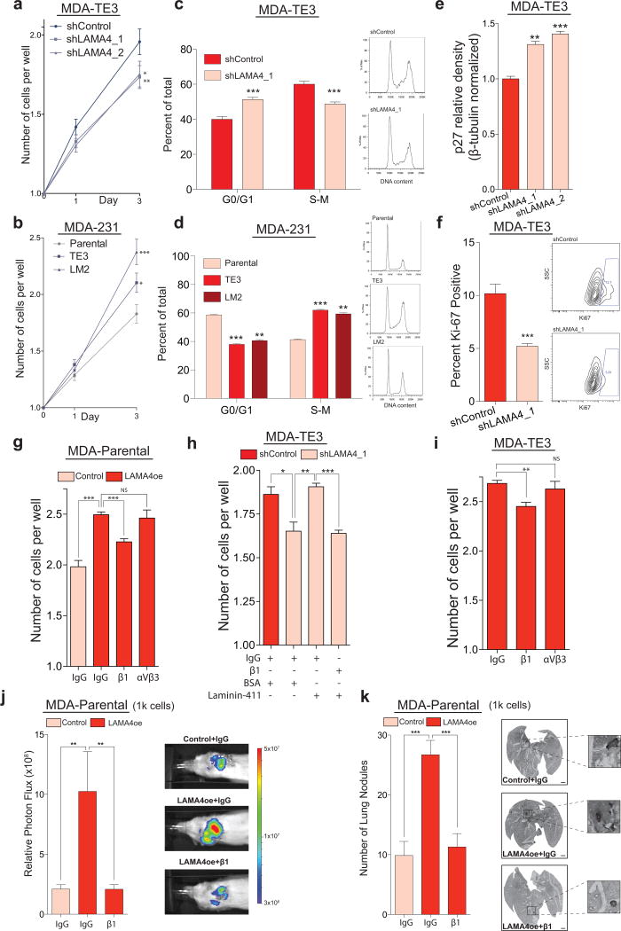
Figure 5. LAMA4 promotes cancer cell proliferation in the absence of substratum-attachment in vitro in a β1-integrin dependent manner.
(a) MDA-TE3 control cells or MDA-TE3 LAMA4-knockdown cells were sorted at clonal density (one cell per well) into low-attachment 96-well plates. LAMA4-depletion led to reduced proliferation. n = 212 (shControl), n = 219 (shLAMA4_1), n = 224 (shLAMA4_2) wells. (b) MDA-parental, MDA-TE3, or MDA-LM2 cells were sorted at clonal density into low-attachment plates. MDA-TE3 and MDA-LM2 cells proliferated more extensively than MDA-parental cells. n = 152 (parental), n = 163 (TE3), n = 141 (LM2) wells. (c) Cell-cycle analysis of MDA-TE3 control cells or MDA-TE3 LAMA4-knockdown cells (left). Representative DNA content histograms (right). n = 6 independent samples. (d) Cell-cycle analysis of MDA-parental, MDA-TE3, or MDA-LM2 cells (left). Representative DNA content histograms (right). n = 4 independent samples. (e) Relative p27 protein levels in MDA-TE3 control cells or MDA-TE3 LAMA4-knockdown cells quantified by Western. p27 values normalized to β-tubulin. n = 3 independent samples. Representative blot in Supplementary Fig. 5l. (f) The fraction of Ki67-positive cells was assessed in MDA-TE3 control cells or MDA-TE3 LAMA4-knockdown cells using flow-cytometry (left). Representative plots (right). n = 6 independent samples. (g) Incubation with β1-integrin blocking antibody suppressed the proliferation of MDA-parental cells over-expressing LAMA4 upon sorting at clonal density into low-attachment plates. Counts on day 3. n = 4 independent experiments. (h) The capacity of recombinant LAMA4-containing protein (laminin-411) to rescue the proliferation of MDA-TE3 LAMA4-knockdown cells was assessed in the presence/absence of β1-integrin blocking antibody upon sorting at clonal density into low-attachment plates. Counts on day 3. n = 3 independent experiments. (i) Incubation with β1-integrin blocking antibody suppressed the proliferation of MDA-TE3 cells upon sorting a clonal density into low-attachment plates. Counts on day 3. n = 3 independent experiments. (j–k) 1×10³ MDA-Parental control cells or LAMA4-over-expressing (LAMA4oe) cells were injected directly into the lung parenchyma in the presence of IgG control or β1-integrin blocking antibody to assess ectopic tumor re-initiation capacity. Lung bioluminescence was measured on day 39 (j). n = 7 independent mice. On day 39 lungs were sectioned, vimentin-stained, and the number of macroscopic nodules per lung was counted (k). n = 7 lungs from 7 independent mice. Scale bars: 1mm. Insets are magnified 5x. *P<0.05, **P<0.01, ***P<0.001 obtained using one-sided Mann-Whitney test (a–b, j–k), or one sided Student’s t-test (c–i). Experiments (a–f) are representative and were replicated at least 2 times. All data are represented as mean +/± S.E.M.

