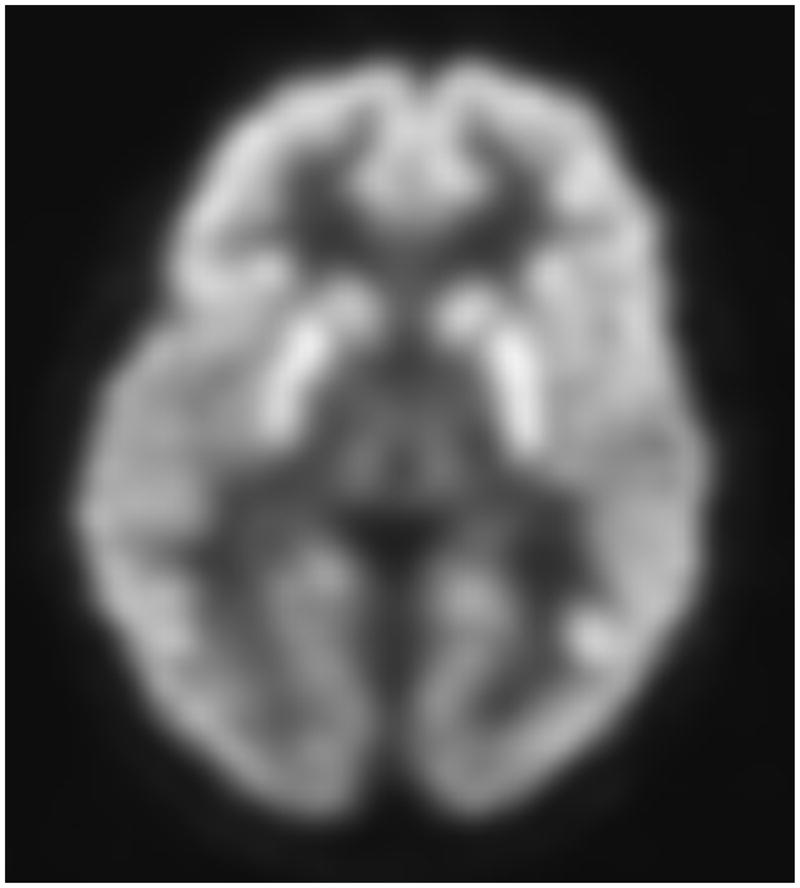
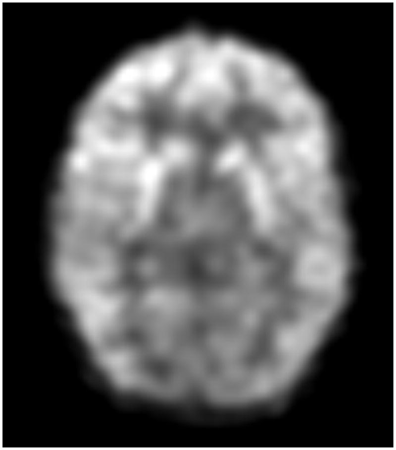
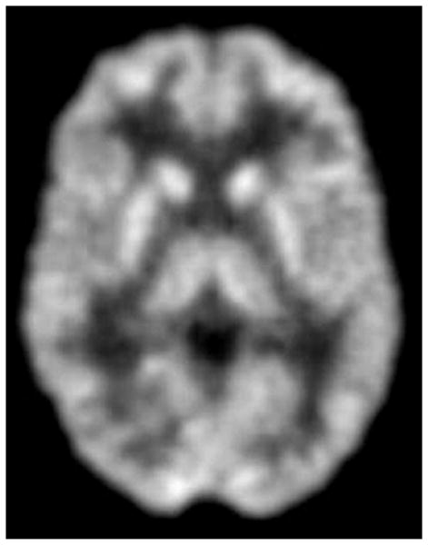
Figure 1.


Transaxial FDG-PET images of a G1D patient of 9 years of age harboring a missense mutation (A) and of a 8 year old G1D patient with no mutation in the coding region of SLC2A1 (B). (C) illustrates a 20-year old normal scan for comparison. The images in A and B are virtually indistinguishable, illustrating the fact that DNA-mutation negative patients are phenocopies (in this and all other respects) of patients genetically diagnosed.
