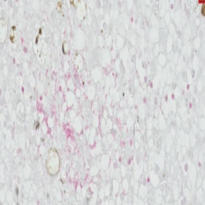
FIG 2.

Gram staining of a section of the liver showing a small number of localized areas highlighting Gram-negative bacteria morphologically compatible with S. enterica (scale bar, 20 μm; magnification, ×600). This photo was taken with Philips Digital Image System.
