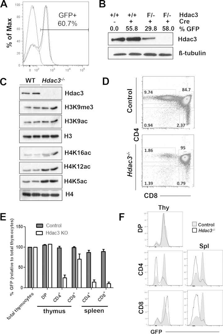
FIG 1.
Loss of Hdac3 affects histone acetylation and thymocyte development. (A) FACS analysis of thymocytes from control mice (no GFP expression) and Lck-Hdac3−/− mice (GFP+). Shown is a tracing for a control mouse that lacks GFP expression and two Lck-Hdac3flox/− mice in that 60% of the thymocytes are GFP+ and thus have the Hdac3 deletion. (B) Western blot of Hdac3 expression in total thymocytes with various degrees of GFP expression. Hdac3 knockout is variable, as indicated by GFP expression. +/+, Lck-Hdac3+/+ mice; F/−, Lck-Hdac3flox/− mice. (C) Western blot of histone modification on thymocytes of either WT-Lck-Hdac3+/+ or Lck-Hdac3flox/− mice sorted using GFP expression. (D) FACS analysis using anti-CD4 and anti-CD8. Unless otherwise noted, plots are representative of those for at least 5 mice. The numbers indicate the percentages of cells analyzed. (E) Bar graphs showing the percentage of GFP-positive T cells from thymic and splenic subpopulations in control or Hdac3−/− mice. Student's t test was used to evaluate each pair, and the differences in CD4+ and CD8+ cells from the spleen were significant (P < 0.05), whereas only the difference for CD4+ cells from the thymus were statistically significant at this level. KO, knockout. (F) FACS analysis of T cells containing CD4 and CD8 versus GFP from the thymus (Thy) and spleen (Spl) showing that Hdac3−/− thymocytes do not persist in the spleen. Plots are representative of those from analyses of at least 5 mice.

