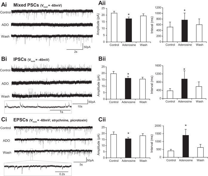
Fig. 2.
Adenosine reduces the frequency and amplitude of both excitatory and inhibitory inputs to interneurons. Ai: voltage-clamp recordings from an interneuron held at holding potential (Vhold) of −60 mV revealing spontaneous synaptic inputs in control, adenosine (ADO), and wash conditions. Aii: averaged data showing that application of adenosine caused a reduction in the amplitude of synaptic events and an increase in the interval between events. Note: these synaptic events represent a mixture of excitatory (EPSCs) and inhibitory (IPSCs) postsynaptic currents (both are depolarizing at this potential in our recording solutions). Bi: voltage-clamp recordings taken from ventral horn interneurons held at −40 mV, a potential at which IPSCs are hyperpolarizing and therefore distinguishable from EPSCs. Bii: averaged data showing that application of adenosine led to an irreversible reduction in IPSC amplitude and a reversible increase in interval between IPSCs. Ci: voltage-clamp recordings of EPSCs taken at −60 mV while inhibitory transmission was blocked with 1 μM strychnine (glycine receptor antagonist) and 60 μM picrotoxin (GABA receptor antagonist). Cii: averaged data showing that application of adenosine led to a reversible reduction in EPSC amplitude and a reversible increase in interval between EPSCs. *Significantly different from control.

