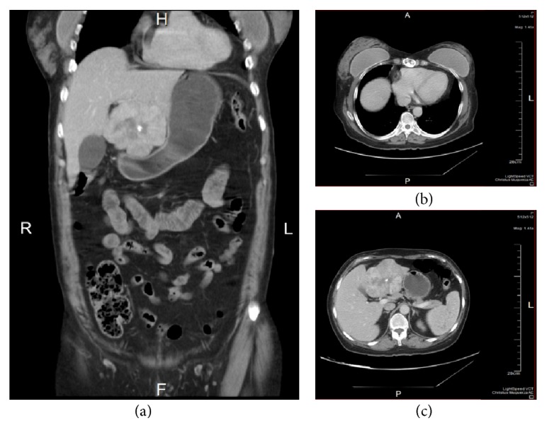
Figure 2.

Images from the abdominal CT. (a) and (b) show the 8 × 8 × 7.3 cm tumor located in segment IV of the left lobe of the liver that laterally displaces the gallbladder. The lesion has defined lobular borders that show a heterogeneous peripheral areas and a hypodense center with calcification. (c) shows changes in the sternum with presence of surgical material.
