Figure 5.
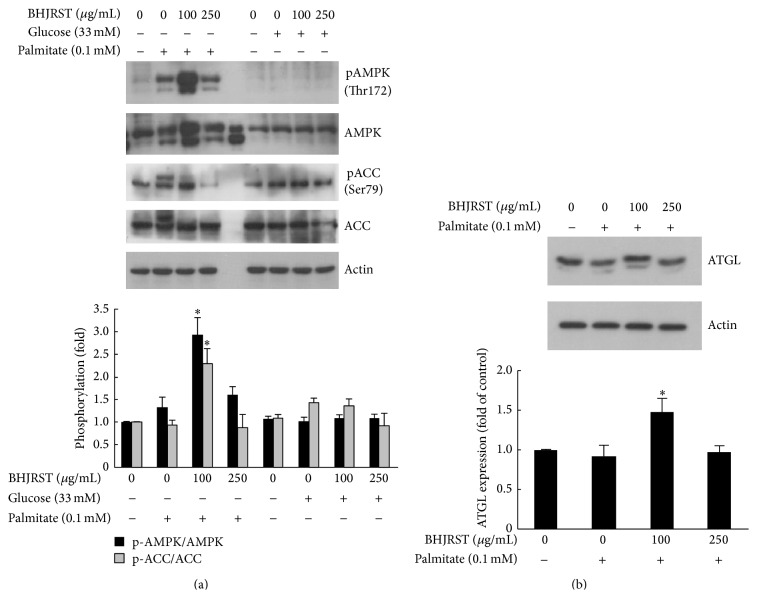
BHJRST stimulates AMPK phosphorylation and ATGL expression under high fat conditions. (a) HuS-E/2 cells were left untreated or were incubated for 24 h with or without 0.1 mM palmitate in glucose-free PH medium with or without BHJRST (100 or 250 μg/mL) (left section) or in PH medium containing 33 mM glucose with or without BHJRST (100 or 250 μg/mL) (right section); then, they were analyzed for phosphorylation of AMPK at Thr172 and ACC at Ser-79, total AMPK and ACC, and actin. Representative immunoblots are shown in the upper panel and the densitometric analysis of AMPK and ACC phosphorylation is shown in the lower panel; the results are the mean ± SD for three independent experiments for the intensity of the phosphorylated band divided by that for the “total” band expressed as a fold value of the control value. (b) Western blot analysis of the expression of ATGL and actin in untreated HuS-E/2 cells and cells incubated for 24 h with 0.1 mM palmitate and 0, 100, or 250 μg/mL of BHJRST as above. The upper panel shows a representative blot and the lower panel shows the quantitative analysis of ATGL expression normalized to actin levels and expressed as a fold value compared to the control value. The data represent the mean ± SD for three independent experiments. ∗ p < 0.05 versus control.

