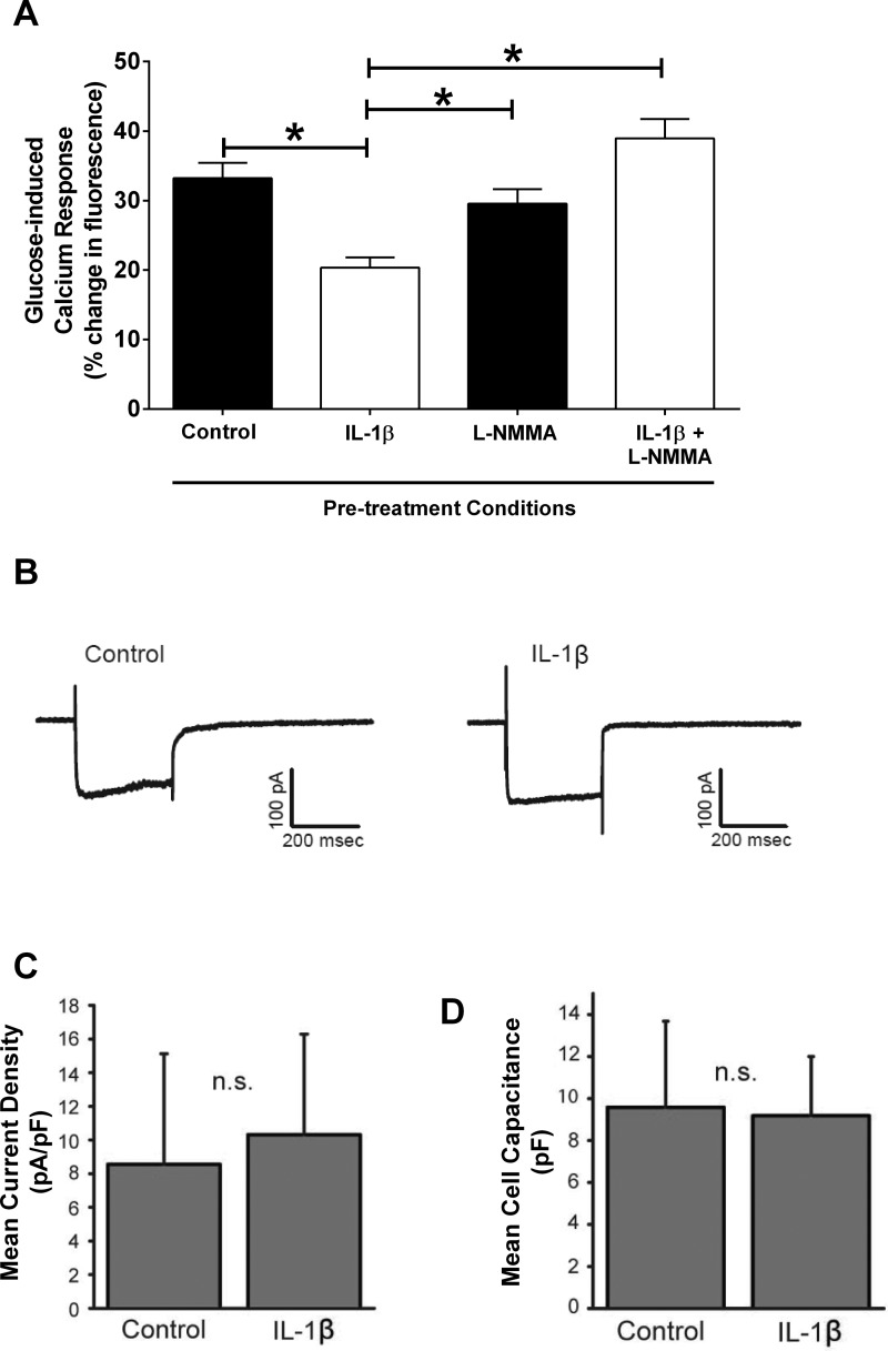
Fig. 6.
Nitric oxide mediates the IL-1β-mediated impairment in glucose-stimulated calcium responses. A: 832/13 cells were cotreated for 18 h with 1 ng/ml IL-1β and 1 mM l-NMMA, followed by a bath-applied glucose challenge to evoke elevations in intracellular calcium levels as measured by changes in fluorescence. *P < 0.05. B: whole cell voltage clamp recordings from a control and an IL-1β-treated cell. Cells were held at −70 mV and then stepped for 300 ms to a series of voltages ranging from −103 mV to +17 mV. Shown are the currents generated by the step to −3 mV. Voltage step values are corrected for the liquid junction potential (−13 mV). C: mean current densities (voltage step to −3 mV) were plotted for control and IL-1β-treated cells. No significant difference in current amplitude was observed (n = 25 for both). D: capacitance values for the 2 groups of cells indicate that cell size for IL-1β-treated cells was also not significantly different from control (n = 25 for both).

