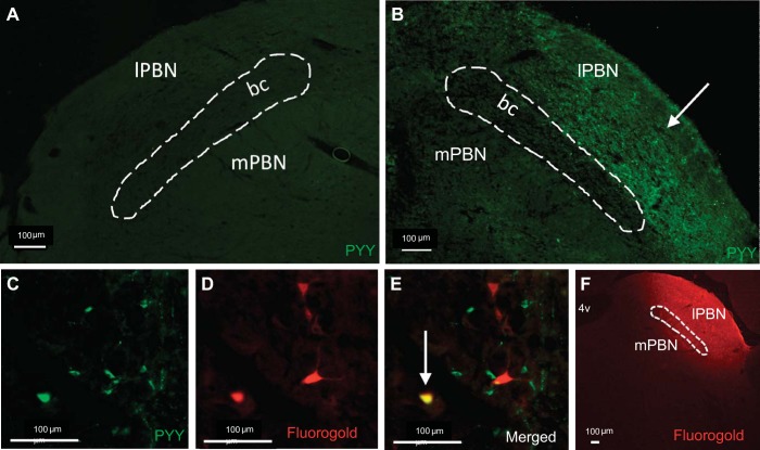
Fig. 2.
A: omission of peptide YY (PYY) primary antibody (negative control) resulted in no PYY fiber immunofluorescence in the lPBN. B: PYY immunohistochemistry confirmed the presence of PYY fibers (white arrow) in the lPBN. C–E: representative ×20 magnification image of a Gi section: (C) green immunofluorescence represents PYY, (D) red immunofluorescence represents lPBN-injected Fluorogold(FG), (E) merged image shows colocalization of FG and PYY (white arrow). F: representative image of lPBN FG injection.

